Expand Your Universe
Ophthalmic Ultrasound Like You've Never Seen Before
Beyond Imaging.
Insightful Analytical Tools.
Explore Our Lineup
VuMAX HD
The gold standard in ophthalmic ultrasound, the VuMAX HD is cutting edge technology providing unparalelled image quality with elegant and powerful usabiity. Simply the best.
B | U | A | diagA
VuPad
Innovation you can see and touch. Use on tabletop or attach to VESA mount arm. Truly outstanding image quality and elegant user interface.
B | U | A | P | diagA
PacScan Plus
Portable, digital, combination A-scan and optional pachymeter, with a large color touch screen, excellent accuracy, repeatability, and reliability.
A | P
Master-Vu
USB probes and exclusive Sonomed Escalon software to turn your laptop computer into an A-scan or B-scan.
B | A
Ultrasound Interconnectivity
From the ability to review images across a clinic from an exam room to the OR to automatically transferring patient and exam data to and from an EHR to save time and prevent errors, the interconnectivity between imaging devices and other clinic systems is increasingly of great importance. There is no one universal way of transmitting data so Sonomed Escalon ultrasound devices provide the widest array of options to ensure our clients can unleash the Power of Communication.


Immersion
A-Scan
Eliminate corneal compression and get accurate measurements – watch this video for instruction in performing immersion A-scan technique

Welcome to the LEO Family
The Leo family of systems consists of the VuMAX HD, VuPad, Master-Vu A systems, which all utilize the same base powerful Leo ophthalmic ultrasound software platform developed by Sonomed Escalon. Specific features and modalities vary between systems but all take advantage of the Leo software for their core functionality, including:

High Resolution Imaging with Enhanced Focus Rendering
Comprehensive Interconnectivity Functionality for Interfacing with PACS, EHRs, and Other Systems
Fast, Robust Performance with Efficient and Configurable Workflow
Advanced Analytical Tools and Features
TM
The Leo family of systems provides the very best image quality, optimized workflow, expansive analysis tools, and unparalleled interconnectivity.
The Importance of UBM
in ICL Sizing
Proper pre-operative selection of ICL size is imperative to avoid risks of excessive vault, elevated IOP and glaucoma, displacement, recurrent uveitis, or anterior subcapsular cataract formation and subsequent explantation, unnecessary risks, and unhappy patients. And UBM is absolutely essential to proper ICL sizing.
ICLguru
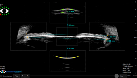
ICLguru is an advanced AI-powered ICL sizing calculator that accurately predicts the post-surgical lens vault and angle for each ICL size available, utilizing next-generation modeling and image analysis of well-aligned high-resolution UBM images to make automated measurements of several parameters for use in its sophisticated nomogram. The information is presented in an easily-interpreted graphical and tabular interface that helps surgeons select the right lens size the first time, every time. Sonomed Escalon UBM AI tools for eye tracking, align assist, and auto capture enable surgeons to obtain optimized UBM scans and achieve accurate results.

SULCUS-TO-SULCUS
Use of sulcus-to-sulcus measurements obtained by UBM input into well-established nomograms has been used by many leading ICL surgeons to achieve significantly improved sizing reliability vs. the OCOS method based on white-to-white. Align Assist and other tools to ensure well-aligned images are exclusively available in the VuMAX HD and VuPad systems to ensure accurate sulcus-to-sulcus measurements and sizing results.
Sonomed Escalon recognizes the significant contributions of Robert Rivera, MD (1956 – 2015) and continues to make available the results of his extensive research and experience as a means of advancing the field of ophthalmology. His presence will be missed, but his legacy will live on.


